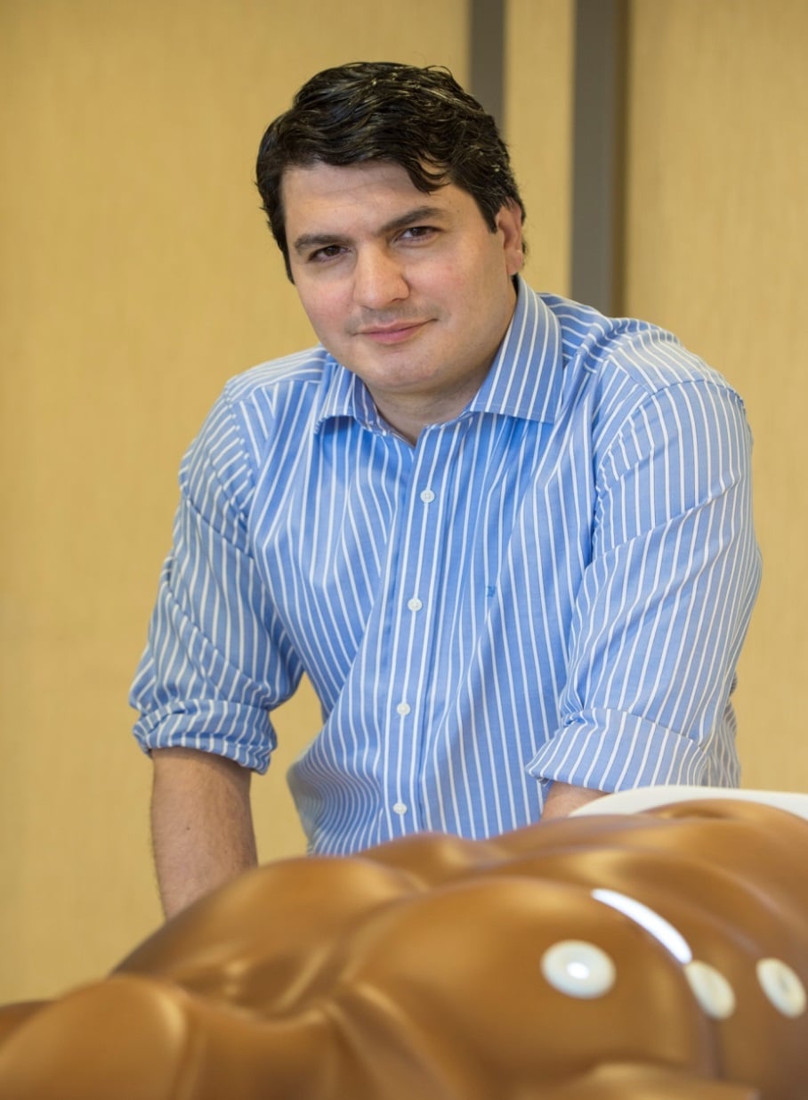
PIEK continues its series of articles on the impact of the electronic interconnect industry on healthcare. This time we talked to Dr Sardari Nia, cardiothoracic surgeon at the Maastricht UMC+, the academic hospital in Maastricht. He was born in Iran, grew up in the Netherlands and studied in Antwerp, Belgium. His fields of activity are invasive valve surgery, mitral valve surgery, robot surgery (MIDCAB) and lung surgery. He seems to be just an ordinary surgeon, but during our talk we notice how much of a technical expert Dr. Sardari Nia is, not only making use of developments in the electronic interconnect industry but even extending and enhancing the cooperation between healthcare and technology together with engineers and technicians for the benefit of his patients.
In order to understand the progress in the field of heart and lung diseases and their treatments, we have to travel back briefly in medical history. Dr. Sardari Nia explains:
 ‘traditionally, there were modes of treatments in health care for all patients with a certain disease, or in other words, one treatment was applied to numerous patients. Now, however, a paradigm shift has taken place. We now believe that personalised healthcare works much better. The focus is now on the individual differences within a certain disease. In practice this boils down to seeing which particular treatment works best for which particular patient. Progress in technology has made this new approach possible.’
To a layman this may seem logical and not very exciting. Let us therefore make clear what far-reaching effects technological innovations can have on heart and lung health care.
If a patient had to be operated on his heart and/or a lung, it used to involve a dramatic, invasive operation in which the breastbone had to be opened completely to get to the heart. The new minimally invasive operation method has radically changed this, because now the surgeon is able to operate from the patient’s sides. This is especially important for replacing or repairing mitral valves in the heart.
Which technological developments have helped to make this possible? Dr. Sardari Nia explains that specialist sets of instruments are required for minimally invasive operations. ‘With the instruments available so far this would not have been an option, but technological progress has yielded ever refined and improved instruments. Furthermore, developments in echoes and CT scans have made great contributions. The images are sharper and of a higher resolution, so that doctors can now even see depths. On top of this, there is 3D echocardiography and computer tomography. These technologies help to virtually reconstruct a patient’s anatomy. What happens is that the inside of a patient’s upper body is reconstructed and that repair options / treatment options are chosen and an operation plan is made on the basis of such a reconstruction. The prime aim is to make as few cuts as possible, so to minimize the risks for the patient.’
Dr. Sardari Nia goes one step beyond in marrying healthcare with technology. Here too we have to remember that every patient’s anatomy is different. This is essential when repairing or inserting mitral valves. After the organs, blood vessels, valves, etc. in the upper body have been virtually recreated, 3D print technology is used to make a mock-up of the mitral valve for a patient. This is an individualised mock mitral valve for one particular patient. These methods ensure increased patient safety and risk minimization. In fact, surgeons are first practising in a mock environment before they actually touch the patient.
In order to practise as much as possible and to optimize operation methods Dr. Sardari Nia has developed a simulator in cooperation with technicians and engineers, the so-called ‘high-fidelity endoscopic mitral valve repair simulator’. It is a construction made of plastics in the shape of an upper body, but without the head and arms. On the sides there are various apertures used to simulate an endoscopic operation or actual operation with the help of robot surgery.
When asked by PIEK what the advantages are of the simulator, Dr Sardari Nia gives the following explanation: ‘In the USA about five operations are executed per surgeon involving mitral valves every year. It is logical that a surgeon cannot gain extensive experience, let alone routine, due to this low number of operations. Here too, practice makes perfect. By using the simulator endless practising and experience building has suddenly become possible. This has led many surgeons from all over the world to travel to the academic hospital UMC+ in Maastricht to practise with the simulator under the supervision of Dr Sardari Nia.
At the end of our interview we ask Dr. Sardari Nia to list the advantages of the synergy between technology and healthcare. ‘The three main effects,’ he says, ‘that I see of using technological innovations in healthcare are:
1. Increased patient safety
2. Higher effectiveness of treatments
3. The reproducibility of operations
All three effects reinforce each other and help us to provide better care.’
Finally, PIEK asked in which countries the companies can be found that help him. ‘These companies can be found all over the world,’ he comments, ‘they are innovative companies that understand how important it is that doctors and engineers work together. Traditionally technology and healthcare were separate rather than connected worlds, but these companies have adopted a mind-set in which the two are inextricably linked. The two worlds are no longer separate, but they are one.’
That is an inspiring end to an extremely interesting talk.